Written By | Health News Agencies
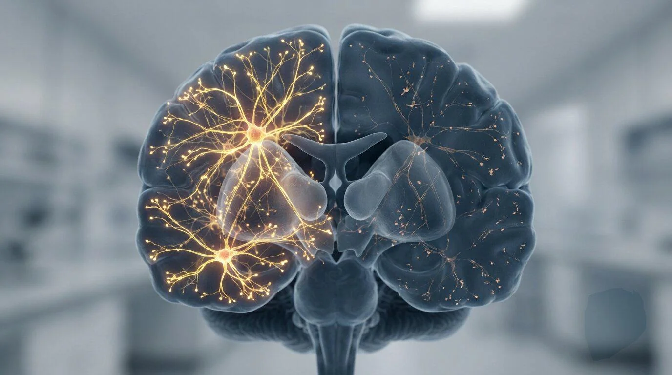
Everybody loses brain tissue with age — but not everybody loses their sharpness.
HEALTH
Every family knows the puzzle, even if nobody in it has ever put words to the question.
Two people, same age, same town, same rough hand of cards. Both of them shrinking a little each year in the way that all human brains shrink. One of them, at eighty, is still doing the crossword in pen, still telling stories with every name in the right place, still winning arguments. The other has started reaching for words that used to come free.
Scan them both and the scans might look almost identical. Same grey matter loss. Same thinning cortex. Same unhelpful verdict from the machine.
So what separates them?
Scientists at the Mark and Mary Stevens Neuroimaging and Informatics Institute at the Keck School of Medicine of USC believe they have found at least part of the answer — and it is sitting in a layer of the brain that most research has been quietly ignoring for decades.
Their findings were published in Alzheimer's & Dementia: The Journal of the Alzheimer's Association, and they have put a name to a kind of hidden armour: superficial white matter.
THE LOCAL ROADS OF THE HUMAN BRAIN
To understand what the researchers were looking at, picture the brain as a country.
Grey matter is the cities. It sits on the outside of the brain, a wrinkled outer layer packed with the nerve cell bodies that do the actual work of thinking — recognising a face, retrieving a word, deciding whether to cross the road.
White matter is the road network. It is made of the long fibres that carry signals from one place to another, wrapped in a fatty insulation called myelin that gives it its pale colour.
Most brain research has concentrated on the motorways: the thick, long-distance bundles running deep through the brain, connecting front to back and left to right. Those are easy to see and easy to measure.
Superficial white matter is different. It is a thin layer of nerve fibres sitting directly beneath the grey matter — short, curved connections running between neighbouring patches of cortex. Not motorways. Local roads. The lane that takes you from one part of a town to the part next door.
And because the fibres are short, curved and crammed right up against the grey matter, they are genuinely difficult to image. For most of the history of brain scanning, they have been the part of the map left blank.
"Gray matter and superficial white matter are physically close and may play different roles: gray matter processes information, while superficial white matter helps nearby brain regions communicate," said Yingxu Liu, PhD, a postdoctoral scholar at the Stevens INI and the study's first author.
"Our findings suggest that cognitive health depends not only on how much gray matter is preserved, but also on the condition of the wiring that connects it."
459 PEOPLE, AND A KIND OF SCANNER THAT SEES WATER MOVE
The study brought together brain imaging and detailed cognitive testing from 459 adults aged 60 and over, living in communities across India. To see the invisible layer, the team used an advanced form of diffusion MRI. Rather than photographing tissue directly, this technique tracks the way water molecules move through the brain — and because water moves differently depending on what is in its way, the pattern of that movement can reveal microscopic structure that an ordinary scan cannot.
The researchers focused on two things in particular. The first was neurite density: neurites are the fine projections through which nerve cells send and receive their signals, and a sparser forest of them suggests damage. The second was free water — the amount of fluid sloshing about unconstrained in the tissue. More free water can point to loss of myelin, to inflammation, or to swelling.
Participants were then put through a battery of cognitive assessments covering language, memory, executive function and visuospatial ability.
The clearest signal in the whole dataset was language.
People whose superficial white matter was in better condition performed better on language tests. The strongest associations of all sat in the frontotemporal regions of the brain — the areas that handle recognising words, speaking fluently, and holding language in mind for the few seconds it takes to finish a sentence.
Which, if you have ever watched someone you love grope for a word that will not come, is not an abstract finding at all.
THE FINDING THAT CHANGES THE STORY
Here is where the study stops being interesting and starts being important. Grey matter loss was still, as expected, the strongest single predictor of how well someone performed cognitively. Nobody is claiming otherwise. Losing the thinking tissue matters.
But the researchers found that the damage done by that loss was not fixed. It varied — and it varied according to the health of the superficial white matter sitting right underneath it.
When the local wiring was in poor condition, grey matter loss hit hard: it was much more strongly linked to impaired language and to broader cognitive difficulty.
When the local wiring was healthy, the same amount of grey matter loss did far less visible damage. The link between shrinkage and decline weakened.
In other words: the wiring appears to act as a buffer.
"The findings point to superficial white matter as a possible source of resilience," said Leon Aksman, PhD, assistant professor of research neurology at the Stevens INI and the study's senior author.
"Two people with a similar degree of gray matter loss may not experience the same cognitive effects if the local connections surrounding that gray matter differ in health. Following participants over time will be essential to test whether preserving these connections can help maintain cognition."
That is the puzzle from the top of this article, restated in the language of neuroimaging. Same loss, different outcome — and now, for the first time, a candidate explanation you can actually measure on a scanner.
WHY THIS STUDY WAS DONE IN INDIA — AND WHY THAT MATTERS ENORMOUSLY
The data came from LASI-DAD, the Harmonized Diagnostic Assessment of Dementia for the Longitudinal Aging Study in India. It is the first nationally representative study of dementia in the country, drawn from a parent survey of more than 72,000 adults aged 45 and over across 36 states and union territories. The scale of what it is measuring is sobering. Wave 1 of LASI-DAD estimated dementia prevalence among Indians aged 60 and over at 7.4 per cent — which translates to something in the region of 8.8 million people living with dementia in India alone.
But the reason this study matters beyond its own borders is who is in it. More than half of the wider LASI-DAD population has low literacy. About 60 per cent live in rural communities. These are precisely the people who almost never appear in brain imaging research, which has historically been built on volunteers who are wealthier, more educated, more urban and overwhelmingly drawn from high-income countries.
This work is among the first to examine superficial white matter in a community-based population from a low- and middle-income country.
And the pattern it found was not uniform. The association between superficial white matter and language ability was stronger among people who could not read or who read incorrectly, among those who had received no formal education, and among participants living in rural areas.
The researchers are careful — pointedly careful — about how far that can be pushed. The study does not show that illiteracy or rural living caused changes in brain tissue. What it suggests is that brain ageing is shaped by a tangle of lifelong influences: education, social circumstance, general health, and environmental exposure, all interacting in ways a single scan cannot untangle.
"A fuller understanding of brain aging requires research that reflects the world's social, cultural, and geographic diversity," said Arthur W. Toga, PhD, director of the Stevens INI and Provost Professor at USC.
"By studying an underrepresented population and looking beyond gray matter alone, this work brings us closer to identifying the biological and social factors that may protect cognition across the lifespan."
WHAT THIS MEANS FOR YOU
Here is the honest answer, and it is worth reading carefully before anybody starts selling you something on the back of this research. There is currently no scan you can book, no supplement you can buy, and no exercise you can do that has been proven to thicken or repair your superficial white matter. Anyone who tells you otherwise is well ahead of the evidence.
What this study does is shift the framing of a question that millions of people quietly ask themselves in their sixties and seventies. For years, the mental model of brain ageing has been essentially one of loss — how much tissue have I got left, and how fast is it going?
This work suggests the more useful question may be about connection. Not just how much grey matter survives, but how well what remains can still talk to itself.
That reframing is not merely philosophical. It points research towards a different target. White matter is known to be sensitive to things that are, at least in principle, modifiable: vascular health, blood pressure, inflammation, metabolic disease. If superficial white matter turns out to be protective and it turns out to respond to those factors, then the everyday, unglamorous business of controlling blood pressure and looking after your blood vessels acquires an entirely new significance for the mind as well as the heart.
None of that is established. All of it is now testable — which is more than could be said a week ago.
There is also a quieter message here about who gets studied. If the brain-ageing map has been drawn almost entirely from educated, urban, high-income volunteers, then the conclusions built on it may simply not describe most of the people ageing on this planet. This study is one attempt to redraw that map.
THE LIMITS — AND THEY ARE REAL
The single biggest constraint is timing. Every participant was examined at one point only. That makes this a snapshot, and a snapshot cannot tell you what came first.
Researchers do not yet know whether superficial white matter starts to deteriorate before grey matter loss begins, alongside it, or ahead of any cognitive decline that can be measured. All three are possible. The study cannot distinguish between them.
Nor can it establish cause. It is entirely conceivable that some third factor — vascular disease, say, or chronic inflammation — damages both the wiring and the thinking tissue, and that the apparent buffering effect is a reflection of that shared cause rather than a protective mechanism in its own right.
The authors say plainly that long-term research following people as they age will be needed to sort this out. They have already flagged the next questions: how vascular health, inflammation, Alzheimer's-related proteins and other biological factors interact with changes in grey and white matter together.
It is also worth noting that this is a study of association across a population, not a prediction about any individual. A healthy-looking scan is not a guarantee, and a poor-looking one is not a sentence.
The work was supported by the US National Institute on Aging, the National Institute of Mental Health, the National Institute of Neurological Disorders and Stroke, and the Office of the Director of the National Institutes of Health.
THE LAYER NOBODY WAS LOOKING AT
Science has a habit of finding its answers in the places it previously found too awkward to look. Superficial white matter has been sitting there in every brain scan ever taken — a thin band of short, curly fibres too fiddly to measure properly, quietly doing the local work of letting one part of the mind speak to the part next door.
Now that the imaging has caught up with it, it turns out that the band may be carrying a good deal more weight than anyone gave it credit for.
It will take years of follow-up to know whether protecting it is possible, let alone how. But for anyone who has ever looked at two people the same age and wondered why one of them is still so sharp, there is now a serious scientific answer forming — and it is not about how much brain you have left.
It is about whether the parts you still have can reach each other.