Scientists built separate ageing clocks for male and female organs and found something nobody expected: the same brain scan carries a completely different warning for a woman than it does for a man. In one measure, her risk more than doubles.
HEALTH
Two people walk into a scanner. Same age on the birth certificate — sixty-eight, say. Same scan, same machine, same afternoon. The images come out looking, to an untrained eye, more or less alike.
For decades the algorithms that read those images treated the pair as interchangeable. Feed in the brain, get back a number: your brain looks four years older than you are. Same number, same meaning, whoever you are.
A paper published last week in Nature Medicine says that assumption has been quietly wrong, and that the cost of it lands hardest on women.
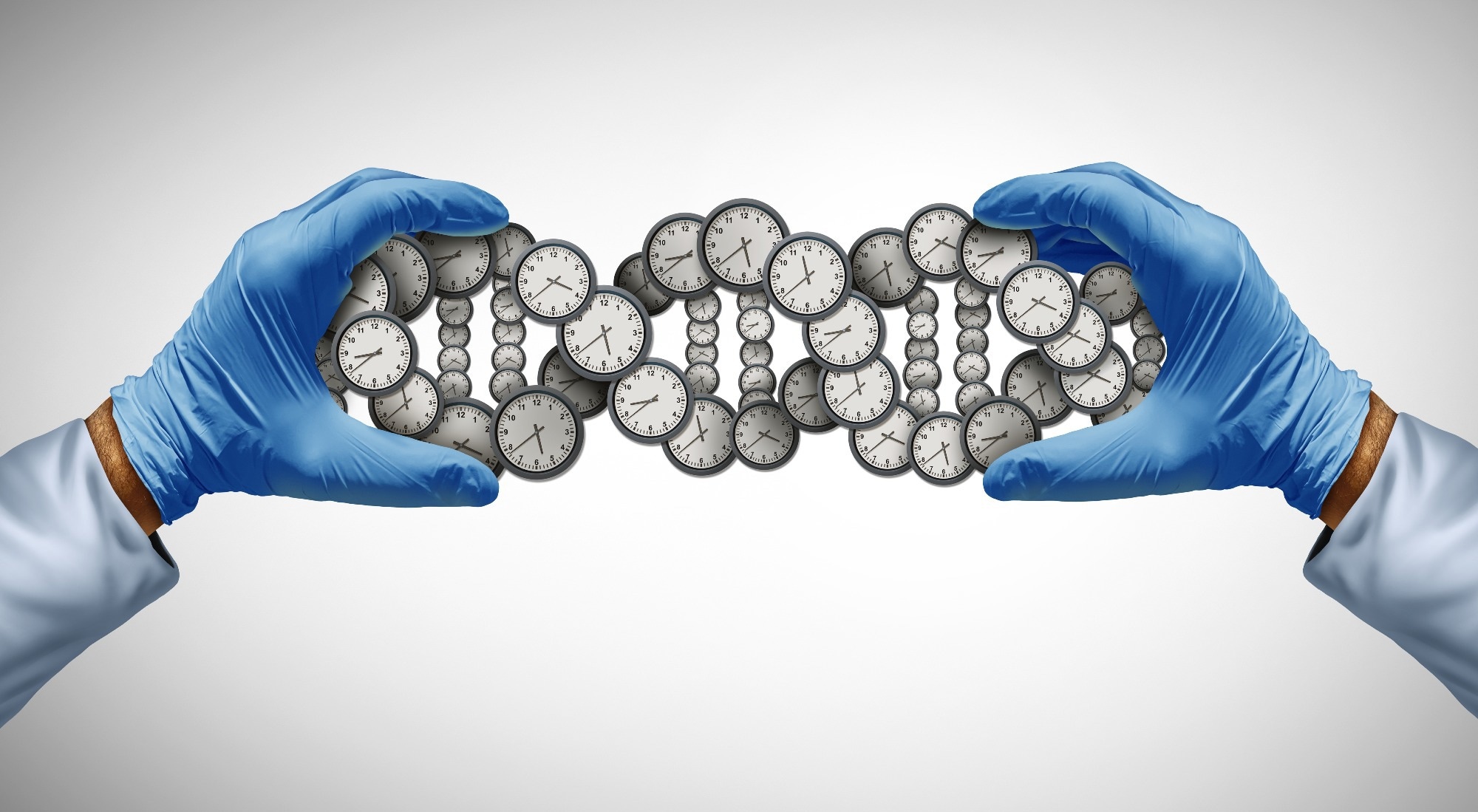
Researchers working under the banner of the MULTI Consortium built not one ageing clock but thirty-eight of them — one set for males, one for females, spanning fifteen organ systems. Then they ran the same brain measurement through both. In men, a brain that looked one standard deviation older than expected came with a 74 per cent higher risk of converting from mild cognitive impairment to Alzheimer's disease. In women, the risk more than doubled.
Same scan. Same number on the readout. Two entirely different futures.
WHAT A BIOLOGICAL AGE GAP ACTUALLY IS
Strip away the jargon and the concept is one most people already understand intuitively. Everybody knows a sixty-year-old who could pass for forty-five and a forty-five-year-old who has clearly had a hard paper round.
Biological age puts a number on that intuition. An algorithm is trained on thousands of healthy people to predict chronological age from some measurable feature — the shape and volume of brain tissue on an MRI, the concentration of particular proteins in blood, the profile of small molecules in the bloodstream. Once trained, you feed it a new person. If it guesses sixty-five for someone who is fifty-eight, that seven-year surplus is the biological age gap, and it is the surplus, not the prediction, that carries the information.
The MULTI team built three flavours. MRIBAGs from imaging. ProtBAGs from plasma proteins. MetBAGs from metabolites. Multiply those across fifteen organ systems and split each one by sex, and you arrive at thirty-eight clocks.
The data came from an unusually deep well: genetic, proteomic, metabolomic, MRI and longitudinal clinical records curated by the consortium, with the UK Biobank supplying the genome-wide association work and roughly two decades of hospital inpatient follow-up. The Alzheimer's Disease Neuroimaging Initiative supplied the dementia-progression data. The Baltimore Longitudinal Study of Aging was used to replicate the protein findings independently. Disease genetics came from FinnGen and the Psychiatric Genomics Consortium.
And then, critically, the clocks were tested against a real randomised drug trial — the A4 study of solanezumab in people with preclinical Alzheimer's.
THE SPLEEN THAT TELLS ON MEN
The single most striking finding is not about the brain at all. It is about the spleen — an organ most people could not locate on themselves.
When the researchers asked which blood proteins track with spleen ageing, the answer in men was forty-six proteins, including tumour necrosis factor, one of the central alarm bells of the inflammatory system.
In women, the same analysis returned four. Just four: ITGBL1, FASLG, MFGE8 and NCR1, all immune-modulating.
Forty-six versus four, in the same organ, from the same kind of blood sample. A sex-blind model averages those two signals together and produces something that describes neither person.
The brain showed the same asymmetry in miniature. In women, one protein stood out — SLITRK1, associated with a younger-looking brain. In men, eight did, including GDF15, ADAM22 and SEZ6L.
Even the heritability differed. Liver and metabolic ageing measures were more strongly inherited in women. Immune, spleen and skin measures were more strongly inherited in men. Endocrine protein ageing was markedly more polygenic in women — spread across many more genetic variants — while immune protein ageing showed the reverse.
These are not rounding errors. They are different biological architectures wearing the same organ names.
DIFFERENT ORGANS, DIFFERENT DISEASES
Because the team had two decades of hospital records to test against, they could ask the question that actually matters: does an ageing clock predict what goes wrong later?
It does — but not the same thing in both sexes.
In women, an accelerated heart MRI clock predicted future hypertension. An accelerated immune metabolic clock predicted high cholesterol. Accelerated digestive and immune metabolic clocks predicted type 2 diabetes. Liver and digestive metabolic ageing showed the strongest links, supported by Mendelian randomisation, to cardiometabolic outcomes.
In men, the dominant signals came from the endocrine and metabolic clocks, and they pointed to high cholesterol, hypertension, diabetes, lipoprotein abnormalities, coronary interventions and statin use.
On death itself, the split persisted. Twelve clock-mortality pairings reached significance in women, driven by protein clocks across multiple organs plus the metabolic and immune metabolic clocks. In men there were fourteen, with a broader risk pattern — and, curiously, the spleen and liver MRI clocks appeared to run the other way, looking mildly protective.
On the metabolic clock specifically, the strongest female signals were for Alzheimer's disease, sleep disturbances and psychiatric traits. The strongest male signals were for metabolic traits and diabetes-related microvascular damage.
Alzheimer's, sleep and psychiatric illness in one column. Microvascular damage in the other. Same measurement.
THE DEMENTIA NUMBERS
The brain findings are where the paper stops being interesting and starts being uncomfortable.
Roughly two-thirds of Americans living with Alzheimer's are women. The long-standing explanation is that women live longer and age is the dominant risk factor — which is true, and probably not the whole story.
The MULTI clocks predicted progression from mild cognitive impairment to Alzheimer's in both sexes. But the strength differed sharply. In men, a one standard deviation increase in brain age carried a hazard ratio of 1.74. In women, 2.25.
Worth noting precisely what the clocks did not do: they did not significantly predict the earlier transition, from cognitively normal to mild cognitive impairment, in either sex. This is a tool for people already showing symptoms, not a crystal ball for the healthy.
Then there is the trial result, which is the part likely to cause arguments at conferences for the next year.
Among participants in the A4 trial who received solanezumab, women whose brains were ageing more slowly than expected scored better on the preclinical Alzheimer's cognitive composite at week 240 — about four and a half years after randomisation — than men in the same category. Among participants with accelerated brain ageing, that sex difference vanished.
Put plainly: in this analysis, biological brain age appeared to separate who responded and who did not, and it did so differently for women than for men. If that holds up, it is a finding about how anti-amyloid drugs get tested, not merely how ageing gets measured. Solanezumab itself failed to slow decline in A4 — but a trial that cannot distinguish between the people in it will struggle to detect a benefit even where one exists.
WHAT THE AUTHORS THEMSELVES SAY IS WRONG WITH IT
Credit where it is due: the limitations section of this paper is unusually honest, and any responsible account has to give it equal billing.
The genetic analyses were restricted to people of European ancestry. That is a serious constraint on a paper about population differences, and it means the findings cannot be assumed to travel to Black, Asian, Hispanic or mixed-ancestry populations without being tested there first.
Many of the disease genetics pulled from FinnGen and the Psychiatric Genomics Consortium were themselves sex-pooled — the very thing the paper argues against — which blunts some of the comparisons.
The UK Biobank has limited repeat imaging and repeat omics data, so most of this is a snapshot rather than a film. The authors flag within-person trajectories as future work precisely because they could not do it here.
The models did not incorporate hormonal status, pregnancy history, or the social and gender-related exposures that shape health over a lifetime. For a paper about sex differences, that is a substantial hole, and the authors say so. Menopause alone reorganises cardiovascular, skeletal and metabolic risk in ways no protein panel in this study accounted for.
Age prediction performance was, in the authors' own word, modest. External validation at biobank scale has not yet happened.
And the concluding recommendation is measured rather than triumphant. The divergence between male and female clocks supports sex-stratified modelling when the goal is a sex-specific normative reference — but sex-pooled and sex-interaction analyses remain complementary approaches, not discarded ones. This is a paper arguing for an addition to the toolkit, not a replacement of it.
THE LONGER ARGUMENT THIS FEEDS INTO
There is history behind this. Women were routinely excluded from early-phase clinical research in the United States until guidance changed in the 1990s, on a rationale that mixed protecting potential pregnancies with a frank desire to avoid the statistical nuisance of the menstrual cycle. The consequence was decades of dosing and diagnostic thresholds derived largely from male bodies and applied to everyone.
The medical consequences are well documented. Women having heart attacks are more likely to present with symptoms that do not match the textbook and more likely to be sent home. Autoimmune disease overwhelmingly affects women and remains comparatively under-explained. Adverse drug reactions occur more frequently in women, a pattern that has been linked in part to dosing derived from male pharmacokinetics.
What the MULTI paper adds is that the problem does not stop at the clinic. It reaches into the measuring instruments themselves. If your ageing clock was trained on a pooled population, it is not neutral — it is a weighted average that fits the majority contributor best and describes everyone else slightly wrongly.
And an unhelpfully averaged clock is not a harmless one. These models are already being used to select patients for trials, to stratify risk, and — increasingly — to sell consumer biological-age tests.
WHAT THIS MEANS FOR YOU
Let us be direct about the practical implications, because the gap between what this study shows and what a marketing department could claim it shows is enormous.
This is not a test you can ask for. There is no sex-specific organ ageing panel at your doctor's office, and there will not be one soon. The authors explicitly say external validation has not been done.
If you have bought a consumer biological age test, treat the number with scepticism. Most commercial clocks are trained on pooled data and most report a single headline age for your whole body. This paper's central finding is that both of those design choices lose information — and that the loss is not evenly distributed.
If you are a woman with a family history of dementia, the honest takeaway is uncomfortable but limited. The study suggests brain ageing may carry more predictive weight in women once mild cognitive impairment is already present. It says nothing useful about a healthy woman's odds. It is not a reason to seek a scan, and a scan would not currently be interpreted this way in any case.
What is actionable is the same unglamorous list it always is, because the things that move these clocks are the things that move every other measure of cardiometabolic health: blood pressure control, sleep, physical activity, not smoking, and managing cholesterol and blood sugar. The organ clocks that predicted trouble in this study — heart, liver, immune, metabolic — are precisely the ones responsive to those levers.
And if you are of non-European ancestry, be aware that you were largely not in this dataset. That is not a small footnote. It is the difference between a finding that applies to you and one that has not yet been tested on you.
THE BOTTOM LINE
Thirty-eight clocks is, on one level, an act of methodological housekeeping — a demonstration that a modelling shortcut the field has used for years discards real signal.
But housekeeping is how medicine actually improves. The finding that forty-six proteins track spleen ageing in men and four do in women is not a curiosity. It is evidence that when researchers pooled the sexes to gain statistical power, they were averaging away the thing they were trying to measure.
The gap between a hazard ratio of 1.74 and one of 2.25 is the difference between a warning and an alarm. For years the same scan delivered the same sentence to both. On the strength of this paper, it should not have.
Whether that translates into anything a patient can use depends on work that has not been done yet: validation in other biobanks, in other ancestries, with hormonal history included, tracking the same people over time rather than freezing them at one moment.
Until then, the honest headline is narrower than the exciting one. Not that scientists can now tell you how fast you are ageing. Only that they have proved they were previously telling half the population something meant for the other half.
Published By | Features Editor